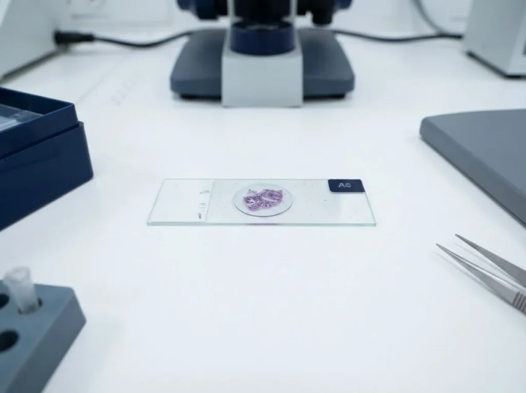
A standardized procedure for mounting non-germinating microscopy specimens for optical examination. Each step protects the slide, the specimen, and the clarity of the field of view.
Sanitize the slide, deposit a small sample, place the cover slip at an angle, then perfuse stain from one edge.
1. Sanitize the Slide
Wipe the glass slide with a lint-free tissue to remove residue and particulate before mounting.
2. Deposit the Sample
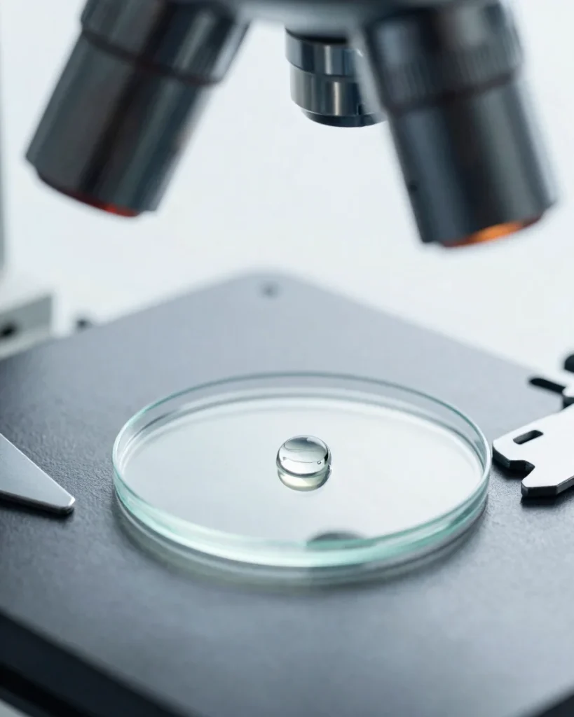
Place a small, measured aliquot of the suspension at the centre of the slide to limit spreading.
3. Angle the Cover Slip
Lower the cover slip at a shallow angle, touching the drop first, to reduce trapped air bubbles.
4. Perfuse the Stain
Apply stain at one edge of the cover slip and draw it across with filter paper on the opposite edge.
Optical Technique
Immersion Oil and Slide Handling
For high-magnification objectives, a single drop of immersion oil bridges the cover slip and the lens front element, matching refractive index and improving resolution of the specimen.
Handle slides by their edges only, keep cover slips clean, and wipe oil from the objective after each session to prevent contamination of subsequent preparations.